
Cell culture
COLO320DM, COLO320HSR and SNU-16 cells (ATCC CCL-220, CCL-220.1 and CRL-5974, respectively) were maintained in RPMI-1640 medium supplemented with 10% fetal bovine serum (FBS), 100 U ml−1 penicillin-streptomycin, 1% non-essential amino acids, and 2 mM l-glutamine. HEK293T cells (ATCC CRL-3216), PC3-DM, and PC3-HSR (gifts from the P. Mischel laboratory) were cultured in Dulbecco’s Modified Eagle Medium (DMEM) supplemented with 10% bovine calf serum, 100 U ml−1 penicillin-streptomycin, 1% non-essential amino acids, and 2 mM l-glutamine. NCI-H716 cells (ATCC CCL-251) were cultured in RPMI-1640 with the same supplements. All cells were incubated at 37 °C in a humidified atmosphere containing 5% CO2. All cells were tested for Mycoplasma by PCR.
HeLa cells were cultured as previously described7. In brief, cells were grown in DMEM supplemented with 10% FBS (Omega), 100 U ml−1 penicillin, 100 U ml−1 streptomycin, and 2 mM l-glutamine. For MTX experiments, dialysed FBS was used. Specific HeLa-derived clones were used at the following MTX concentrations: PD29424h (H, DM+, 60 nM MTX) and PD29426a (HSR+, 40 nM MTX).
Adult neuronal stem cells (aNSCs) from Mycec/+; Trp53fl/fl (ecMyc) or control Myc+/+; Trp53fl/fl (control) mice were isolated and cultured in adherent condition on laminin-coated (Sigma-Aldrich, L2020) dishes in NeuroCult Stem Cell Basal Media with NeuroCult Proliferation Supplement (Mouse & Rat) (Stem Cells Technologies, 05702), 20 ng ml−1 EGF (Stem Cells Technologies, 78006), 10 ng ml−1 bFGF (Stem Cells Technologies, 78003), and 2 μg ml−1 heparin (Stem Cell Technologies), ecMyc and control aNSCs were treated by Adeno-Cre (from ViraQuest (VQ-Ad-CMV-Cre; 1 × 10¹² particles per ml; 091317)) to induce engineered ecDNA formation in ecMyc cells. Cells were maintained in culture for 3 weeks to accumulate Myc-containing ecDNAs. All cells were negative for mycoplasma contamination. Cells were maintained in a humidified 5% CO2 atmosphere at 37 °C.
For proliferation assays, cells were seeded at clonal density in 12-well plates. The following day, cells were treated with a drug (Polθ inhibitors, MTX, CHK1 inhibitor or FGFR inhibitor). For HeLa experiments, MTX concentrations were applied as described above. In both the HeLa and SNU-16 experiments, cells were pre-treated with Polθi or vehicle for 1 week prior to replating for growth assessment. Medium with fresh drug was replaced every 72 h. Cell proliferation was monitored using the IncuCyte Live-Cell Imaging System (Essen BioScience/Sartorius), with confluency measurements collected every 24 h.
For proliferation assays involving FANCM loss and Polθ inhibition, FANCM-knockout cells were generated using the triple-guide CRISPR–Cas9 strategy described below, with AAVS1-targeted cells serving as controls. Two days after knockout, cells were plated at 2,000 cells per well in 48-well plates in three technical replicates for each condition, and Polθi-Pol treatment was initiated. Cell proliferation was monitored using the IncuCyte Live-Cell Imaging System with confluence measurements acquired every 12 h.
Drug treatments
The following compounds were used in this study: Polθ inhibitors: RP-2119 (Polθi-Hel; Repare Therapeutics; 100 nM), RP-6685 (Polθi-Pol; Repare Therapeutics; 10 µM); DNA-PKcs inhibitors: NU7441 (Selleck Chemicals, S2638; 1 µM), NU7026 (Selleck Chemicals, S2893; 10 μM); CHK1 inhibitor (CCT245737; Selleck Chemicals, S8253; 750 nM); methotrexate (MTX; Sigma-Aldrich, 454126; concentrations varied by cell line and clone as noted above); hydroxyurea (Sigma-Aldrich, H8627; 100 μM); PARP inhibitor (Olaparib; Selleck Chemicals, S1060; 1 μM); thymidine (Sigma-Aldrich, T1895; 2 mM); nocodazole (Sigma-Aldrich, M1404; 100 ng ml−1); FGFR inhibitor (Infigratinib/BJ398; Selleck Chemicals, S2183; 6.25 nM); triptolide (Millipore, 645900; 1 µM); and CDK9 inhibitor (AZD4573; Selleck Chemicals, S8719; 200 nM).
For FISH experiments and proliferation assays, the media were replaced every 72 h with fresh medium containing the indicated drugs. Drug concentrations were maintained consistently throughout the assay.
CRISPR–Cas9 ribonucleoprotein complex assembly and nucleofection
CRISPR RNA (crRNA) and trans-activating CRISPR RNA (tracrRNA) (Integrated DNA Technologies, IDT) were each resuspended in IDTE buffer to a final concentration of 100 µM. Equimolar amounts were mixed to yield a 50 µM crRNA:tracrRNA (sgRNA) duplex, which was annealed by heating to 95 °C for 5 min, followed by cooling to room temperature. For ribonuclear protein (RNP) complex formation, purified Cas9 protein (Berkeley Facility; and sgRNA were mixed at a 1:1.2 molar ratio and incubated at room temperature for 20 min before use. In experiments using three sgRNAs simultaneously to knock out a gene, sgRNAs were designed to be between 35 and 300 bp apart. Each sgRNA was resuspended in IDTE buffer and pooled to a final concentration of 40 µM per guide. RNP complexes were assembled by mixing 104 pmol of Cas9 with 120 pmol of the combined sgRNA mix (1:1.15 molar ratio) and incubating at room temperature for 20 min. Target cells were prepared using the SF Cell Line 4D-Nucleofector X Kit (Lonza) according to the manufacturer’s instructions. Nucleofection was performed using the Lonza 4D-Nucleofector System with program CM158. Knockouts were validated by PCR amplifying the region around the sgRNA target site, followed by Sanger sequencing and TIDE49 or ICE CRISPR Analysis (EditCo Bio., v3.0). For simultaneous RHINO and FANCM knockouts, a triple-guide CRISPR–Cas9 strategy was used for each gene, and RNP complexes targeting both genes were combined at a 1:1 ratio before nucleofection. Cells were resuspended in SF solution, mixed with the combined RNP complexes, and nucleofected as described above. For the corresponding single-knockout controls, IL25-targeting RNPs were included to match the total guide load in the double-knockout condition. Sequences for sgRNA in Supplementary Table 1. PCR primers for TIDE/EditCo ICE and CRISPR analysis in Supplementary Table 2.
Lentiviral production and transduction
Lentivirus was produced in HEK293T cells using a standard four-plasmid transfection system. For each transfection, 5 µg of RRE, 3 µg of VSV-G, 2.5 µg of REV, and 20.5 µg of a BFP-expressing BRCA2 shRNA transfer plasmid were combined with 62 µg ml−1 polyethylenimine (PEI) and 150 mM NaCl. The DNA-PEI complexes were incubated at room temperature for 15 min before being added to 10-cm dishes of HEK293T cells at ~70–80% confluence. Cells were incubated overnight at 37 °C in a humidified 5% CO2 incubator. The following day, the culture medium was replaced, and after a 6–8 h recovery period, the first viral supernatant was collected. This process was repeated twice at 24-h intervals to collect the second and third viral supernatants. Fresh medium was added after each collection.
The BRCA2 knockdown construct targeted exon 16 of the BRCA2 coding sequence and was designed using the Broad Institute’s RNAi design tool. The hairpin sequence (5′-GCGTTTCTAAACATTGCATAA-3′) was cloned into the pLKO.1-puro vector (Addgene #8453). Target cells were transduced with the lentiviral supernatant and selected with puromycin to generate stable knockdown lines. Functional validation of BRCA2 depletion was assessed by measuring cell proliferation in response to 1 µM olaparib. Cell growth was monitored using the IncuCyte Live-Cell Imaging System (Essen BioScience/Sartorius), with confluency measurements recorded every 24 h.
Generation of RHNO1
−/− cells
COLO320DM cells were stably infected with pLentiCRISPRV2 Cas9 – blast (Addgene #98293) and selected with blasticidin (5 μg ml−1). Blasticidin-resistant cells were then stably infected with pLenti-GFP-2A-Puro-gRHINO1_A (Addgene #211613) and selected with puromycin at 3 μg ml−1. RHNO1 knockout was confirmed with TIDE analysis (forward: TTTTGCTTGGTGGTTGTAGG, reverse: TATCTGGCATTCTCACCCAG).
Generation of Fucci4 cells
Lentivirus was prepared containing one of each of the following plasmids, gifts from M. Lin, to generate Fucci4 cells50: pLL3.7m-Clover-Geminin(1-110)-IRES-mKO2-Cdt(30-120) (Addgene #83841), pLL3.7m-mTurquoise2-SLBP(18-126)-IRES-H1-mMaroon1 (Addgene #83842), mKO2-SLBP(18-126) (Addgene #83914) and Clover-Geminin(1-110) (Addgene #83915). COLO320DM and COLO320HSR cells were transduced with lentiviral supernatant containing either mTurquoise2-SLBP/H1-mMaroon1, mKO2-SLBP, or Clover-Geminin. Transgene-positive cells were isolated by FACS and used as colour controls in experiments. A split of mTurquoise2-SLBP/H1-mMaroon1-positive cells was transduced with lentiviral supernatant containing Clover-Geminin/mKO2-Cdt, and transgene-positive, fully labelled Fucci4 cells were isolated by FACS.
Cell cycle analysis of Fucci4 cells by flow cytometry
COLO320DM or COLO320HSR Fucci4 cells were plated and treated for 72 h with either DMSO (0.1–0.3% as required per control), Polθi-Pol, Polθi-Hel, DNA-PKcs inhibitors, Chk1i, Chk1i plus Polθi-Pol, or Chk1i plus Polθi-Hel. Cell cycle controls included a double thymidine block using 2 mM thymidine or an overnight treatment with 100 ng ml−1 nocodazole. For the experiment, CRISPR–Cas9-mediated sgRNA targeting of either AAVS1 or FANCM was performed in COLO320DM Fucci4 cells. Knockout cells were plated and treated for 72 h with either 0.1% DMSO, Polθi-Pol, or Polθi-Hel. Cells were trypsinized, collected in RPMI medium, washed with DPBS, pelleted, resuspended in DPBS with 3% FBS, and filtered using a 35-μm mesh into polystyrene tubes (Falcon; 38030) for flow cytometry. Cells were then analysed on an Aurora full-spectrum analyser (Cytek Biosciences), with 100,000 to 150,000 events collected per condition, and quantification was performed using FlowJo (v10.10.1). Single or dual-colour controls (mTurquoise2–SLBP/H1–mMaroon1, mKO2–SLBP or Clover–Geminin) and unstained cells were used to determine the gating strategy for G1-, S–G2– and M-phase cells (Supplementary Fig. 1).
Generation of H2B–GFP and H2B–RFP cells
Lentivirus was prepared containing one of each of the following plasmids, gifts from E. Fuchs: H2B–GFP (Addgene #25999) and H2B–RFP (Addgene #26001). COLO320DM and COLO320HSR cells were transduced with H2B–GFP and H2B–RFP separately, and transgene-positive cells were isolated by FACS. Labelling was confirmed by live-cell microscopy in 35 mm no. 1.5 glass-bottomed dishes (Mattek; P35G-1.5) using a Nikon Ti2 inverted microscope equipped with a TokaiHIT STX stage-top incubator and objective heating collar (37 °C, 5% CO2), a Yokogawa CSU-W1 spinning disc confocal unit, and dual Hamamatsu C14440-20UP sCMOS cameras (Dual Fusion configuration). The system was controlled using NIS-Elements AR software version 6.10.02. Cells were imaged using an Apo TIRF 60× 1.49 NA oil immersion objective (Nikon). Four fluorescence channels were acquired sequentially using laser excitation from an LUN-F multi-laser unit and 300 ms exposures. Excitation wavelengths and laser powers were as follows: 405 nm (100 mW) at 30% power, 488 nm (100 mW) at 10% power, and 561 nm (100 mW) at 30% power. A quad-band dichroic mirror (Di01-T405/488/568/647) was used with the following emission filters: 455/50 nm (DAPI), 526/36 nm (GFP), and 605/52 nm (dsRed–mCherry), split across two detection paths using a 561 nm long-pass dichroic (DM A561LP). Images were saved in Nikon ND2 format and subsequently processed using Fiji (ImageJ version 2.16.0)51.
Fluorescence-based competition assays
CRISPR–Cas9-mediated targeting of IL25 or POLQ (see ‘CRISPR–Cas9 ribonucleoprotein complex assembly and nucleofection’) was performed in COLO320DM and COLO320HSR cells labelled with either H2B–GFP or H2B–RFP. Each cell type was mixed 1:1 in the following pairings and plated in triplicate at 2,000 cells per well in a 48-well tissue culture plate (Corning Costar; 3548):
|
H2B label |
Knockout |
H2B label |
Knockout |
|---|---|---|---|
|
RFP |
sgIL25 |
GFP |
sgPOLQ |
|
RFP |
sgIL25 |
GFP |
sgIL25 |
|
RFP |
sgPOLQ |
GFP |
sgIL25 |
1:1 mixtures were imaged on an IncuCyte Live-Cell Imaging System (Essen BioScience/Sartorius) for up to 2 weeks. Knockouts were confirmed during the experiments using DNA extraction with the DirectPCR Lysis Reagent (Viagen; 301-C), PCR amplification of the knockout regions, Sanger sequencing and EditCo’s ICE Analysis tool (v3). Cell confluency, the number of GFP-positive cells, and the number of RFP-positive cells were quantified using the IncuCyte Analysis software. Technical replicates were averaged, and the number of RFP- or GFP-positive cells per condition was normalized first to timepoint 0 (day 1), and then to the H2B–RFP (sgIL25) or H2B–GFP (sgIL25) control condition.
Live-cell imaging of mitotic timing
COLO320DM or COLO320HSR cells expressing H2B–GFP were plated on 35 mm no. 1.5 glass-bottomed dishes (Mattek; P35G-1.5) and treated for 48 h with either 0.1% DMSO or Polθi-Hel. At 48 h, cell medium was changed to RPMI without phenol red (supplemented with respective drugs or treatments). Time-lapse images were acquired on a Nikon Ti2 inverted microscope equipped with a TokaiHIT STX stage-top incubator and objective heating collar (37 °C, 5% CO2), a Lumencor Sola II light engine, and a Hamamatsu C14440-20UP sCMOS camera (SN:000740). The system was controlled using NIS-Elements AR software version 5.21.03. Cells were imaged using a Plan Apo λ 100× 1.45 NA oil immersion objective (Nikon). Two fluorescence channels were acquired at each time point: GFP (ET-GFP; Nikon; 96366; ET470/40X; ET525/50 M; 200 ms exposure) and DIC. Time-lapse imaging was performed with a 3-min interval between frames, and the Perfect Focus System (PFS) was engaged. Images were saved in Nikon ND2 format and subsequently processed in Fiji (ImageJ version 2.16.0)51 to manually screen for mitotic events.
Polθ ATPase enzymatic assay
In a 384-well assay plate (Corning 3572), recombinant full-length human POLQ (3 nM) was combined with a 10-point concentration range of Polθi-Hel (highest concentration 0.1 mM, threefold serial dilutions) in a buffer containing 50 mM Tris-Cl pH 7.5, 10% glycerol, 5 mM DTT, 10 mM MgCl2, and 0.1 mg ml−1 BSA. The plate was incubated at room temperature for 15 min, after which the reaction was initiated by adding a DNA substrate (20 nM) and ATP (100 µM). Plates were incubated for 90 min at room temperature, and ATPase activity was subsequently detected using an ADP-Glo assay kit (Promega) according to the manufacturer’s protocol. In brief, 15 μl of ADP-Glo reagent was added to each well, followed by a 1 h incubation at room temperature. 30 μl of Detection reagent was then added per well, the plates were incubated for 45 min at room temperature, and luminescence was read on an Envision plate reader (Revvity). The forked DNA substrate was prepared as follows: oligos 1–3 were mixed (1:1:1) in a buffer containing 10 mM Tris-HCl, pH 7.5, 50 mM NaCl, and 1 mM EDTA, heated to 95 °C for 5 min, and cooled to room temperature.
Oligo 1: 5′-GCACTGGCCGTCGTTTTACGGTCGTGACTGGGAAAACCCTGGCG-3′; oligo 2: 5′-TTTTTTTTTTTTTTTTTTTTTTCCAAGTAAAACGACGGCCAGTGC-3′; oligo 3: 5′-TTGGAAAAAAAAAAAAAAAAAAAAAA-3′.
Expression and purification of recombinant full-length Polθ
Four 1-l cultures of 293-6E cells were grown in F17 medium supplemented with 0.1% Pluronic F-68, 4 mM GlutaMAX, and 25 μg ml−1 G418 in 2-l shake flasks. Cultures were transfected with 1 mg l−1 of the phCMV1-2m6h-POLQ plasmid (lab of S. Doublié) using PolyPlus PEIPro at a 1:1 (w/v) DNA:PEI ratio. Transfected cultures were maintained at 37 °C with 5% CO2 and shaken at 135 rpm. Cells were collected 4 days post-transfection by centrifugation at 1,000g for 5 min at 4 °C. For lysis, the resulting cell pellet was resuspended in 250 ml of lysis buffer (20 mM Tris-HCl, 250 mM NaCl, 250 mM KCl, 0.01% NP-40, 10% glycerol, pH 8.0) supplemented with 5 mM β-mercaptoethanol and EDTA-free protease inhibitors (Roche). Cells were lysed by freeze–thaw cycles followed by processing through a microfluidizer. The lysate was stirred at 4 °C for 30 min, then clarified by centrifugation at 20,000g for 60 min at 4 °C.
The soluble fraction was batch-bound to 5 ml of amylose resin (New England Biolabs) in buffer A (20 mM Tris-HCl, 200 mM NaCl, 200 mM KCl, 0.01% NP-40, 10% glycerol, 5 mM β-mercaptoethanol, pH 8.0) for 1.5 h at 4 °C with gentle nutation. The resin was first collected in a 5-cm Econo-column, transferred to a 2.5-cm column, and washed with 30 column volumes of buffer A. Elution was carried out using buffer B (buffer A supplemented with 50 mM maltose). The eluate was diluted threefold with pre-heparin dilution buffer (10 mM HEPES, pH 7.5), then loaded at 2 ml min−1 onto a 5 ml HiTrap Heparin Sepharose HP column (Cytiva, 17040703) pre-equilibrated in buffer D (20 mM Tris-HCl, 500 mM NaCl, 500 mM KCl, 0.01% NP-40, 5% glycerol, 5 mM β-mercaptoethanol, pH 8.0). After loading, the column was washed with 20 column volumes of buffer C (20 mM Tris-HCl, 100 mM NaCl, 0.01% NP-40, 5% glycerol, 5 mM β-mercaptoethanol, pH 8.0), and 5 ml fractions were collected. The protein was eluted using a linear 0–100% gradient of buffer D over 20 column volumes at 2 ml min−1. The peak fractions (heparin pool 1) were concentrated to ~0.8 ml using a Vivaspin6 centrifugal device (10 kDa MWCO, PES membrane), then subjected to size-exclusion chromatography using a Superose 6 Increase column (Cytiva) equilibrated in destination buffer (20 mM Tris-HCl, 200 mM NaCl, 100 mM KCl, 2% glycerol, 3 μM sucrose monolaurate, 0.5 mM TCEP, pH 8.0). Fractions (0.5 ml) were collected over 1.1 column volumes and analysed by SDS–PAGE. Protein-containing fractions were pooled and concentrated using a Vivaspin6 centrifugal device (10 kDa MWCO, PES membrane) for downstream applications.
Metaphase FISH for detection of MYC, FGFR2 and DHFR ecDNA
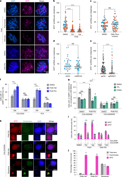
Cells were seeded to reach approximately 50% confluence 36–48 h before collection. To enrich for mitotic cells, Colcemid (0.1 µg ml−1) was added for 6 h, followed by trypsinization. Cells were pelleted by centrifugation (5 min at 1,000 rpm), gently resuspended in 5 ml of pre-warmed 0.075 M KCl, and incubated at 37 °C for 15–30 min with occasional mixing to induce swelling. After centrifugation, residual KCl was retained, and fixative (3:1 methanol:acetic acid) was added dropwise with gentle mixing. The total volume was adjusted to 10 ml with fixative, and samples were stored at 4 °C overnight or longer. For slide preparation, fixed cells were pelleted and resuspended in a minimal volume of fresh fixative. Cold, wet microscope slides were prepared by briefly soaking in water, and cells were dropped from a height onto tilted slides. Slides were immediately placed on a humidified 80 °C heating block for 1 min to promote chromosome spreading, then air-dried overnight. Slide quality was assessed by light microscopy.
To detect extrachromosomal MYC, FISH was performed using a locus-specific probe targeting the MYC genomic region (Empire Genomics: MYC FISH probe). Slides were rehydrated in PBS for 5 min, fixed in 4% formaldehyde for 2 min, and washed 3 times in PBS (5 min each), followed by three washes in 2× SSC (5 min each). Slides were then denatured in 70% formamide/2× SSC at 72 °C for 2 min, immediately dehydrated through an ethanol series (70%, 90% and 100%), and air-dried. The MYC probe, resuspended in hybridization buffer, was applied to the slide, denatured at 80 °C for 5 min, and hybridized overnight at 37 °C in a humidified chamber. The following day, excess probe was removed by sequential washes: 0.4× SSC at 72 °C for 5 min, followed by 2× SSC with 0.05% NP-40 at room temperature. Additional washes were performed in 50% formamide/2× SSC at 42 °C, 2× SSC, and 0.1× SSC. Slides were counterstained with DAPI and mounted using ProLong Gold antifade reagent. Fluorescence microscopy was used to visualize MYC-positive ecDNA, which appeared as distinct fluorescent foci located outside the main chromosomal arms in metaphase spreads.
Detection of additional ecDNA species was performed as follows. For FGFR2, a probe targeting the FGFR2 genomic region (Empire Genomics: FGFR2 FISH probe) was used; FISH was carried out as described above. For DHFR, a probe targeting the DHFR genomic region (Empire Genomics: DHFR FISH probe) was used; FISH was performed using the same protocol.
Detection of MYC-containing micronuclei by interphase FISH
To detect MYC-containing micronuclei, cells were seeded on poly-l-lysine-coated glass coverslips 96 h prior to fixation. Polθ inhibitor or vehicle was added 72 h before collection, followed by hydroxyurea or PBS 24 h before fixation. Cells were fixed in 4% paraformaldehyde (PFA) for 5 min, washed three times with PBS for 5 min each, and permeabilized with 0.5% Triton X-100 in PBS for 10 min. After two additional PBS washes (5 min each), coverslips were rinsed three times in 2× SSC for 5 min and then dehydrated through a graded ethanol series (70%, 90% and 100%). For FISH, the MYC probe (Empire Genomics) was mixed with hybridization buffer, applied to the coverslip, and denatured at 80 °C for 5 min. Hybridization was carried out overnight in a humidified chamber. The following day, coverslips were fixed in freshly prepared methanol: acetic acid (3:1) for 10 min at room temperature, air-dried, and incubated in 2× SSC. Denaturation was performed in 70% formamide/2× SSC at 72 °C for 2 min, followed by rapid dehydration in ethanol (70%, 90%, 100%). The fluorescently labelled MYC probe was then applied and hybridized overnight at 37 °C. The excess probe was removed by post-hybridization washes in 50% formamide/2× SSC at 42 °C, followed by rinses in 2× SSC and 0.1× SSC. A final wash was performed in 0.4× SSC at 72 °C for 5 min, followed by a rinse in 2× SSC containing 0.05% NP-40 at room temperature. Nuclei were counterstained with DAPI and mounted using ProLong Gold antifade reagent. Imaging was performed by fluorescence microscopy. MYC-positive micronuclei were identified as small, DAPI-stained structures that were spatially separated from the primary nucleus and displayed distinct MYC FISH signals.
DNA telomere FISH
Cells (2 × 10⁵) were plated onto poly-l-lysine-treated coverslips and treated with Polθi-Pol, Polθi-Hel, or 0.1% DMSO for 72 h or 0.5 μM Reversine for 24 h, prior to fixation. Cells on coverslips were washed 2 × 5 min with 1× DPBS, then fixed with 4% PFA for 10 min, followed by 3 × 5 min washes in 1× DPBS. Cells were then permeabilized with 0.5% Triton X-100 for 10 min and washed with DPBS, 2 × 5 min each. Coverslips were dehydrated in 70%, 95%, and 100% ethanol for 2 min each, then air-dried. Cells were incubated in 30 μl hybridization buffer (70% formamide, 1 mM Tris-Cl, and Roche blocking reagent at 1 mg ml−1) and 100 nM TelC-Cy3 (PNAbio) on slides marked with a wax pen before denaturation at 80 °C for 5 min. Cells were incubated in the dark at room temperature for 2 h, followed by 4× 15 min formamide washes (70% formamide, 10 mM Tris-Cl in H2O). Cells were washed 3× 10 min in PBST (0.1% Tween-20), with 5 μg ml−1 DAPI added to the second wash. Coverslips were air-dried before being mounted with ProLong Glass antifade.
DNA pan-centromere FISH
Cells (2 × 10⁵) cells were plated onto poly-l-lysine-treated coverslips and treated with Polθi-Pol, Polθi-Hel, or 0.1% DMSO for 72 h or 0.5 μM Reversine for 24 h, prior to fixation. Cells on coverslips were washed 2 × 5 min with 1× DPBS, then fixed with 4% PFA for 10 min, followed by 3 × 5 min washes in 1× DPBS. Cells were then permeabilized with 0.5% Triton-X for 10 min and washed with DPBS, 2 × 5 min each. Coverslips were washed 3 × 5 min in 2× SSC, then dehydrated in 70%, 85% and 100% ethanol for 2 min each, then dried completely. Coverslips were incubated with XCE pan-cen (MetaSystems), diluted 1:5 in hybridization buffer (Empire Genomics), and then sealed with CytoBond (SciGene) before denaturation at 80 °C for 10–15 min. After overnight incubation in a humidified chamber at 37 °C, the CytoBond sealant was removed from the coverslips, which were washed in 0.4× SSC (heated to 72 °C) for 5 min, then washed for 30 s in 2× SSC + 0.05% Tween-20. Coverslips were rinsed briefly with ddH2O, then air-dried, and washed 3× 10 min in DPBS (with 5 μg ml−1 DAPI added to the second wash). Coverslips were air-dried before being mounted with ProLong Glass antifade.
Immunofluorescence for the detection of 53BP1 and gH2AX
Cells were seeded on poly-l-lysine-coated glass coverslips and fixed 72 h later following the indicated treatments. Cells were fixed in 4% PFA for 15 min at room temperature, then permeabilized with 0.5% Triton X-100 for 10 min. Cells were then blocked for 1 h at room temperature using a blocking buffer consisting of 1 mg ml−1 BSA, 3% goat serum, 0.1% Triton X-100, and 1 mM EDTA in PBS (pH 8.0). After blocking, cells were incubated for 90 min at room temperature with primary antibodies diluted in the same blocking buffer: rabbit anti-53BP1 (Novus NB100-304, 1:1,000) and mouse anti-γH2AX (Millipore JBW301, 1:1,000). Following three PBS washes, cells were incubated with Alexa Fluor–conjugated secondary antibodies (donkey anti-rabbit 568 and donkey anti-mouse 488, 1:500, invitrogen) for 30 min at room temperature in the dark. Coverslips were then washed three times with PCB and mounted using ProLong Gold Antifade Mountant with DAPI (Thermo Fisher). DNA damage foci were visualized using fluorescence microscopy (Nikon Si) and quantified based on the number of 53BP1 and γH2AX foci per nucleus.
For immunofluorescence experiments involving transcription inhibition, transcription was acutely inhibited for 3 h with triptolide or CDK9 inhibitor either 3 days after Polθ inhibitor or vehicle pretreatment, or 5 days after CRISPR–Cas9-mediated knockout of FANCM or AAVS1. For immunofluorescence experiments assessing DNA damage following CRISPR–Cas9-mediated targeting of the indicated nucleases and helicases, cells were treated with Polθ inhibitor or vehicle beginning 2 days after nucleofection and collected on day 5. For experiments involving CRISPR–Cas9-mediated co-targeting of RHNO1 and FANCM, cells were nucleofected and collected on day 5 for immunofluorescence.
Combined 53BP1 immunofluorescence and MYC-FISH
Cells were seeded onto glass coverslips and allowed to adhere overnight. The following day, cells were fixed in 4% PFA for 15 min at room temperature, then permeabilized with 0.5% Triton X-100 for 10 min. Cells were then blocked for 1 h at room temperature using a blocking buffer consisting of 1 mg ml−1 BSA, 3% goat serum, 0.1% Triton X-100, and 1 mM EDTA in PBS (pH 8.0). After blocking, cells were incubated for 90 min at room temperature with primary antibody diluted in the same blocking buffer: rabbit anti-53BP1 (Novus NB100-304, 1:1,000). Following three PBS washes, cells were incubated with Alexa Fluor–conjugated secondary antibodies (donkey anti-rabbit 488 1:500, Invitrogen) for 30 min at room temperature in the dark. Coverslips were then washed three times with PBS. Cells were then fixed with 4% PFA at room temperature for 20 min, followed by re-permeabilization using 1× PBS containing 0.7% Triton X-100 and 0.1 M HCl on ice for 10 min. Coverslips were then subjected to acid denaturation using 1.9 M HCl for 30 min at room temperature, followed by washes in PBS and 2× SSC. Coverslips were dehydrated through an ethanol series (70%, 90% and 100%) and air-dried. The MYC probe, resuspended in hybridization buffer, was applied to the coverslip, denatured at 80 °C for 5 min, and hybridized overnight at 37 °C in a humidified chamber. The following day, excess probe was removed by sequential washes: 0.4× SSC at 72 °C for 5 min, followed by 2× SSC with 0.05% NP-40 at room temperature. Additional washes were performed in 50% formamide/2× SSC at 42 °C, 2× SSC, and 0.1× SSC. Slides were counterstained with DAPI and mounted using ProLong Gold antifade reagent.
Colony formation assay
COLO320DM cells were plated at clonal density in triplicate in 6-well plates with 1 μM NU7441 or DMSO. 48 h after plating, cells were then treated with 2 Gy of ionizing radiation. Cells were then allowed to grow for an additional 10 days, after which they were fixed in 4% PFA and stained with crystal violet. Colonies were counted with the Fiji ImageJ Colony Area plugin52.
Immunoblot to detect Cre recombinase expression
Cells were lysed in Laemmli buffer, supplemented with protease and phosphatase inhibitors (cOmplete and EDTA-free Protease Inhibitor cocktail; Roche, COEDTAF-RO and PHOSS-RO). Proteins were separated in NuPage Bis-Tris gels (Invitrogen, NP0322BOX), transferred on a nitrocellulose membrane (0.2μm; Bio-Rad), and blocked by incubation with Intercept (TBS) Blocking buffer (LI-COR: 927-60001). The following primary antibodies were used: anti-Cre recombinase (1:1,000; Cell Signaling, 15036) and anti-α-tubulin (1:5,000; Cell Signaling, 2144). The following secondary antibodies were used: IRDye 800 anti-rabbit (LI-COR, 926-32213) and IRDye 680 anti-mouse (LI-COR, 926-68072). Images were acquired using an Odyssey Imaging System (LI-COR). Full uncropped blots with molecular weight markers are available in the supplementary data (Supplementary Fig. 2).
AAVS1 and MYC Repair-seq
Cells were nucleofected using the Lonza 4D-Nucleofector System with the SF Cell Line Kit- following the manufacturer’s protocol. Cas9 RNP complexes were assembled by incubating purified Cas9 protein (Berkeley Facility; with synthetic sgRNAs (IDT) at a 1:2 molar ratio for 10–15 min at room temperature. sgRNAs were designed to target the AAVS1 locus and exon 2 of the MYC gene. Approximately 1 × 10⁶ cells were nucleofected per condition using program CM158 and collected 72 h post-nucleofection to allow sufficient time for editing events and genomic DNA recovery. Genomic DNA was extracted using the ZymoResearch Quick-DNA Genomic Kit according to the manufacturer’s instructions. DNA concentration and quality were assessed using a Qubit fluorometer and agarose gel electrophoresis. A ~450 bp region surrounding each CRISPR cut site was amplified by PCR using locus-specific primers (primer sequences provided in Supplementary Table 2). PCRs were performed using Taq DNA Polymerase (NEB) with the following cycling conditions: initial denaturation at 95 °C for 3 min; 35 cycles of 95 °C for 15 s, 53 °C for 15 s, 68 °C for 30 s; final extension at 68 °C for 5 min. Amplicons were quantified using Qubit and pooled in equimolar ratios. The IGO facility core of MSKCC prepared Pooled PCR products for sequencing. Libraries were sequenced on the Illumina MiSeq platform using 250 bp paired-end reads (MiSeq v2 PE250 kit). All primer pairs were pre-validated to produce amplicons of similar length and efficiency, ensuring uniform sequencing coverage. Sequence quality was monitored across all samples, and depth uniformity was confirmed during post-run analysis. For experiments evaluating MMEJ, a minimum of 700,000 paired-end reads per sample was obtained. For homology-directed repair quantification, a minimum of 30,000 paired-end reads per sample was achieved. Sequencing reads were demultiplexed and aligned with a reference sequence. The CRISPResso2 toolkit was used to quantify editing outcomes53, characterize insertions and deletions, and reconstruct repair events. For each sample, allele fractions for these events were calculated by counting the number of reads with respective mutational signatures identified by CRISPResso2 and dividing the count by the total reads.
Live-cell imaging of ecDNA tethering
Live-cell imaging to assess ecDNA tethering to mitotic chromosomes was performed using COLO320DM TetO-EGFP cells stably expressing H2B–mCherry and TetR–eGFP. Cells were seeded into four-well chamber slides (ibidi) 48 h prior to imaging and treated with DMSO, Polθi-Pol, or Polθi-Hel for 24 or 72 h. Imaging was conducted in a humidified, 37 °C chamber with 5% CO2 using a Nikon SoRa Spinning Disk Confocal system equipped with a Borealis microadapter, Perfect Focus 4, motorized turret and encoded stage, and a 5-line laser launch (405 nm (100 mW), 445 nm (45 mW), 488 nm (100 mW), 561 nm (80 mW) and 640 nm (75 mW)). A PRIME 95B Monochrome Digital Camera and CFI Apo TIRF 60× 1.49 NA objective lens (W.D. 0.12 mm) were used in super-resolution mode. Laser power was set to 5% with a 600 ms exposure time. Images were acquired using NIS-Elements Advanced Research Software on a dual-Xeon imaging workstation and denoised using the default settings. Time-lapse imaging of mitotic cells was initiated at metaphase or anaphase and continued at 5-min intervals until reformation of daughter nuclei. z-stacks were processed as maximum intensity projections, and image adjustments (brightness and contrast) were performed using Fiji. To quantify cytosolic mis-segregation events, TetR+ ecDNA foci were tracked throughout mitosis; foci excluded from daughter nuclei post-mitosis were classified as mis-segregated ecDNA.
END-seq
To induce a control DSB at the MYC locus, COLO320DM and COLO320HSR cells were transfected with a Cas9 RNP complex targeting the MYC gene. The RNP complex was assembled by mixing purified Cas9 protein (Berkeley MacroLab) with an sgRNA targeting exon 2 of MYC (sequence: GCCGTATTTCTACTGCGACG; IDT), to a final concentration of 20 pmol Cas9 and 25 pmol sgRNA. The mixture was incubated at room temperature for 10–15 min. Cells were nucleofected using the Lonza 4D-Nucleofector System with the SF Cell Line Kit and program CM158. After overnight recovery, cells were treated with either the Polθi-Pol or vehicle control for 72 h, then collected for END-seq. For sgRNA-mediated depletion of AAVS1 or FANCM, cells were nucleofected with the triple-guide knockout strategy described above and treated 72 h later with Polθ-Pol inhibitor or vehicle (DMSO). Cells were collected 72 h after treatment for END-seq. END-seq was performed as previously described54. In brief, COLO320 cells were resuspended in cell suspension buffer (10 mM Tris-HCl, pH 7.2, 50 mM EDTA, 2 mM NaCl) and embedded in 0.75% low-melting-point agarose plugs using the Bio-Rad CHEF Mammalian Genomic DNA Plug Kit. Approximately 5 × 10⁶ cells were used per plug, and two plugs were prepared per condition. Plugs were incubated in freshly prepared lysis buffer (10 mM Tris-HCl, pH 8.0, 50 mM EDTA, 150 mM NaCl, 1% SDS) containing Proteinase K (Qiagen) at 50 °C for 1 h, followed by overnight incubation at 37 °C. After lysis, plugs were rinsed and washed with plug wash buffer (10 mM Tris-HCl, pH 8.0, 50 mM EDTA) and TE buffer (10 mM Tris-HCl, pH 8.0, 1 mM EDTA), followed by treatment with RNase A (Qiagen) at 37 °C for 1 h. Additional washes in plug wash buffer were performed to remove residual enzymes. DNA DSB ends were blunted in-plug using Exonuclease VII and Exonuclease T, A-tailed with Klenow fragment (3′→5′ exo−), and ligated to a biotinylated hairpin adaptor. Plugs were then melted, digested with β-agarase I, and DNA was fragmented by sonication to an average size of ~175 bp. Biotinylated fragments were captured with streptavidin-coated beads, end-repaired, A-tailed, ligated to a second adaptor, treated with USER enzyme, and amplified by PCR with barcoded Illumina TruSeq primers. Libraries were purified with AMPure XP beads, size-selected by gel extraction, quantified, and sequenced on an Illumina platform55.
END-seq analysis
Raw sequencing reads were trimmed using Trimmomatic (v0.39)56. Filtered reads were aligned to the human reference genome (hg19) using Bowtie2 (v2.2.5.1)57. Reads mapping to the mitochondrial genome or ENCODE blacklisted regions were excluded from downstream analysis. BAM files were converted to BED format using the bamtobedfunction in bedtools (v2.31.1)58. The ecDNA region was defined as chromosome 8:127433703–129010006 based on the strong enrichment of END-seq signal across this interval. Peaks were called using MACS2 (v2.2.7.1)59 with the parameters–keep-dup all–nomodel–shift −50–extsize 100 -q 0.001–fe-cutoff 5. Annotations for simple repeats were downloaded from the UCSC Genome Browser and overlaps between repeat elements and END-seq peaks were identified using the intersect function in bedtools60. For visualization and downstream analysis, scaled coverage tracks normalized to reads per million were generated using BAMscale61.
Direct library preparation
COLO320DM cells were used for DLP+ experiments. Four populations were prepared: the parental starting population (T0), a DMSO-treated control population cultured for 2 months, a population cultured in parallel for 2 months in the presence of a Polθi-Hel, and a FANCM-knockout population cultured for 1 month. The FANCM knockout was generated using CRISPR–Cas9-mediated targeting, and knockout efficiency was monitored weekly by genomic DNA extraction, PCR amplification of the targeted cut site, Sanger sequencing, and EditCo’s ICE Analysis tool (v3). Single-cell whole-genome libraries were generated using DLP+, as described previously62. In this approach, individual cells are dispensed into nanowell chips containing preloaded primers and processed directly for single-cell library construction. Cells were subjected to lysis and protease treatment, followed by heat lysis, transposition-based fragmentation and adapter insertion, reaction neutralization and indexed PCR amplification. The resulting single-cell libraries were recovered, pooled, size-selected with AMPure XP beads, and sequenced. Single-cell DNA sequencing data was analysed using the publicly available Mondrian pipeline ( within the Isabl platform63. This included alignment using bwa-mem64 and structural variant calling using DeStruct65. DeStruct was run jointly on all four samples. Analysis was restricted to those structural variants that were identified exclusively in a single sample, as these were thought to be newly arising somatic events. For events outside of the amplicon-implicated chromosomes (6, 8 and 13), only those events with at least 3 supporting reads were included.
Sequence analysis of the breakpoint of rearrangement junctions in human tumours
Focal amplifications in human tumour samples from PCAWG (Pancancer Analysis of Whole Genomes), Hartwig (Hartwig Medical Foundation)41, PedPanCan (tumour samples from St Jude and PBTA), GLASS (Glioma Longitudinal AnalySiS) and CUGA (Chinese Urothelial Carcinoma Genome Atlas), detected and classified by Amplicon Architect and Amplicon Classifier, publicly available through the Amplicon Repository ( were analysed41,42,66,67. Only amplifications from whitelisted primary tumour samples were retained for the analysis.
To identify amplicon junctions occurring within TA repeat regions, junction coordinates were intersected with TA/AT-rich repeats (for example, (TA)n, (TATA)n, etc., including reverse complements), extracted from the UCSC Table Browser RepeatMasker track. The intersection was performed using BEDTools intersect v2.31.1. Each dataset was processed individually with the TA-rich repeat annotation from their corresponding reference genome: hg19 (PCAWG, GLASS) and hg38 (CUGA, PedPanCan).
A two-sided Pearson’s chi-squared test (R base function prop.test v4.3.2) was used to determine whether there is a significant difference between the fraction of junctions overlapping TA repeats on ecDNA vs other focal amplification types.
MYC copy number assay using TaqMan
MYC gene amplification was evaluated using TaqMan copy number variation assays on a QuantStudio 6 Flex real-time PCR system. Myc copy number in mouse neuronal stem cells was assessed using the TaqMan Copy Number Assay (probe Mm00734221_cn) and TaqMan Copy Number Reference Assay (Tfrc, 4458367). MYC copy number in human COLO320(DM and HSR) cells was assessed using the TaqMan Copy Number Assay (probe Hs01764918_cn) and TaqMan Copy Number Reference Assay (RnaseP, 4403326). Genomic DNA was isolated using the Zymo Quick-DNA Miniprep kit and amplified using the TaqPath ProAmp Master Mix (Applied Biosystems, A30865), following the supplier’s instructions. Relative copy number variations were calculated using the ΔΔCt method.
RT–qPCR to detect MYC expression
Total RNA was isolated using the NucleoSpin RNA Clean-up kit (Macherey-Nagel) following the manufacturer’s instructions. Columns were treated with DNase I to degrade genomic DNA. 1 µg of purified RNA was reverse transcribed using the iScript gDNA Clear cDNA Synthesis Kit (Bio-Rad) following the suppliers’ instructions. MYC expression was assessed using a 1:10 dilution of cDNA with PowerUp SYBR Green Master Mix (Applied Biosystems) and standard cycling conditions on a QuantStudio 6 Flex real-time PCR system. MYC expression was normalized to ACTB and quantified using the ΔΔCt method.
RT–qPCR to detect cre recombinase expression
Total RNA was isolated using RNeasy Mini Kit (QIAGEN, 74106) following the manufacturer’s instructions. After treatment with DNAse I (Ambion, AM2222), 0.5 μg of purified RNA was retro-transcribed with random hexamers by using SuperScript IV First-strand System (Invitrogen), cre recombinase expression was assessed using a 1:20 dilution of cDNA with PowerUp SYBR Green Master Mix (Applied Biosystems) and standard cycling conditions on a QuantStudio 6 Flex real-time PCR system. Cre recombinase expression was normalized to GAPDH and quantified using the ΔΔCt method. Quantitative PCR with reverse transcription (RT–qPCR) primers are provided in Supplementary Table 3.
Reporting summary
Further information on research design is available in the Nature Portfolio Reporting Summary linked to this article.








Leave a Reply