
Systematic palaeontology
Dinosauria Owen, 1842 sensu28
Theropoda Marsh, 1881 sensu28
Dromaeosauridae Matthew and Brown, 1922 sensu7
Microraptorinae Senter et al., 20045
Microraptorini (Senter et al., 2004)5
Norellraptor barsboldi gen. et sp. nov.
Holotype
130108-MHGU-F4281 (Figs. 1–3; Supplementary Table 1). The complete skeleton of a microraptorine dinosaur with part of the plumage imprints (Supplementary Fig. 1).
Locality and horizon
Lamadong Town, Jianchang County, western Liaoning, China. Jiufotang Formation, Lower Cretaceous. China.
Etymology
The genus name refers to the American palaeontologist Mark Allen Norell (1957-2025), with Latin word “raptor” (“robber”), a frequently used suffix in dromaeosaurid taxonomy. The species name refers to the Mongolian palaeontologist Rinchen Barsbold (1935-2025). The new taxon name remarks and honours the exceptional contributions by R. Barsbold and M. A. Norell to the study of the bird-like dinosaurs.
Diagnosis
Microraptorine dromaeosaurid with the unique combination of features (autapomorphies marked by asterisk; Supplementary Fig. 2): premaxilla with length of prenarial part half the depth of the subnarial body; large promaxillary fenestra occupying the whole rostral third of the antorbital fossa*, and with long axis inclined 35° caudodorsally; pila promaxillaris narrower than the pila interfenestralis and both less than half the diameter of the maxillary fenestra*; pubis gently curved caudoventrally with small foot having the posterior projection less than the diameter of the pubic shaft*. Differential diagnosis: Norellraptor differs from Changyuraptor in having basally-constricted dentary crowns and in the semilunate carpal barely capping the second metacarpal; from Graciliraptor in having the distal end of metatarsal II subequal in width to metatarsal III; from Microraptor in having the rostralmost maxillary teeth proportionally larger and the first three dentary teeth not procumbent, in the presence of a bulbous distal process of the ulna, in having the arctometatarsalian tarsometatarsus, and in the more extensive fusion between the proximal shafts of metatarsals II to IV; from Hesperonychus and Sinornithosaurus in having a proportionally much smaller pubic foot; from Wulong in the relative size of the promaxillary and maxillary fenestrae in the antorbital fossa; and from Zhongjianosaurus in the proportionally shorter tail and sternum relative to the femur, in the more prominent lateral process of the coracoid, in the proportionally shorter and more gracile forelimb compared to the hindlimb, in the smaller deltopectoral fenestra, in the more moderate caudal inclination of the internal tuberosity of the humerus, in the semilunate carpal more convex proximally and covering most of the proximal surface of the first metacarpal, in the lack of a distinct ventral curvature of the penultimate manual phalanges, in the third manual ungual bearing a proximodorsal lip but being less curved ventrally, in the prominent proximolateral spur on the tarsometatarsus, and in the less elongate penultimate phalanx of the third toe.
Description and comparison
The 57 cm long skeleton is complete and suffered only a moderate disarticulation of the appendicular girdles (Fig. 1a). The histological analysis, complete fusion of the tibiotarsi and tarsometatarsi, and the smooth texture of the long bones support a post-juvenile developmental stage at the time of death29.

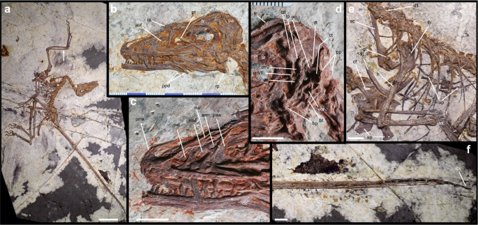
a Whole specimen. b Skull in lateral view. c Close up of the rostrum. d Close up of the posterior part of skull. e Pectoral region. f Distal end of tail. aof, antorbital fossa; arm, ascending ramus of maxilla; at, anterior tympanic recess; bt, basal tuber; bp, basypterygoid process; cf, coracoid fenestra; ct, caudal tympanic recess; cnV, 5th cranial nerve opening; cnVII, 7th cranial nerve opening; cv, cervical vertebra; d1, first dorsal vertebra; de, dentary; dt, dorsal tympanic recess; fo, foramen; fr, frontal; fu, furcula; gl, glenoid; h, humerus; la, lacrimal; lat, laterosphenoid; lcv, last caudal vertebra; lp, lateral pneumatic recess; mc, metacarpus; mxf, maxillary fenestra; na, nasal; oc, otosphenoid crest; or, otic region; pa, parietal; paw, postantral wall; pmx, premaxilla; pp, paroccipital process; ppd, posterior processes of dentary; pt, pterygoid; pxf, promaxillary fenestra; r, radius; rp, retroarticular process; sa, surangular; sc, scapula; so, supraoccipital; sp, sternal plate; sr, sclerotic ring; u, ulna; up, uncinate process; vor, ventral otic recess. Arrow indicates point of histological sampling. Scale bar = 50 mm (a); 10 mm (b–f).
The skull is subtriangular in lateral view, as in other microraptorines9, with a vaulted frontoparietal region and a moderately elongated snout (Fig. 1b-d). The premaxilla is partially preserved and shows a prenarial body proportionally deeper and shorter than in Microraptor and Sinornithosaurus9,30, with the rostral narial margin at the level of the distal end of the second alveolus. The premaxillary teeth are unserrated, as in most paravians. The maxilla is triradiate, similar to other microraptorines9,29,30. The rostral ramus is partially covered by the premaxilla. As in Microraptor9, the caudodorsal ramus of the maxilla is very short, barely extending beyond the level of the ascending ramus. The jugal ramus is triangular, moderately elongate and shallower than the base of the rostral ramus. The depth of the subcutaneous part of the jugal ramus is lower than that of the caudal margin of the antorbital fossa: the two regions are separated by a distinct margin. The antorbital fenestra is “D”-shaped, 150% taller than long. The antorbital fossa is excavated by shallow pits, homologous to the more densely pitted surface in other microraptorines30. The caudal margin of the antorbital fossa is marked by a vertical ridge, which separates the fossa housing the maxillary fenestra from the antorbital fenestra. One diagnostic feature of Norellraptor is the significant fenestration of the maxillary antorbital fossa. The promaxillary fenestra is larger than the maxillary fenestra and occupies the whole rostral third of the antorbital fossa, differing from the much smaller fenestra in Microraptor and Wulong9,29, and from the condition in Sinornithosaurus, whose premaxillary fenestra is larger than the maxillary fenestra but does not occupy the whole rostral third of the antorbital fossa7,30. The maxillary fenestra is enlarged. It is drop-shaped and placed equidistant from the antorbital and the promaxillary fenestrae: accordingly, the pila interfenestralis and promaxillaris are comparably narrow, with the latter narrower than the former and both not wider than half the diameter of the maxillary fenestra. This condition differs from Microraptor and Wulong, which show a much narrower pila interfenestralis and a broad pila interpremaxillaris, and from Sinornithosaurus, which shows both pilae proportionally broader7,9,30. As in Microraptor9, the caudodorsal corner of the maxillary fenestra is not exposed in lateral view, being it roofed by a lip of bone oriented caudoventrally. This feature, absent in other dromaeosaurids31, was originally considered unique of Microraptor, and is here reinterpreted as a synapomorphy of Microraptorini. The lacrimal is “T”-shaped, similar in proportion to other microraptorines9. The postorbital part of the dermal skull bar is eroded, revealing the braincase. The braincase bones are well-preserved and fused to each other (Fig. 1d), differing from the badly preserved condition in other microraptorines9,29,30. The lateral surface of the braincase is extensively pneumatized. An oval pit is present on the laterosphenoid, as in troodontids32,33, and is confluent with the large trigeminal foramen. A sharp otosphenoidal crest separates the dorsal tympanic recess from the otic depression. The lateral surface of the prootic-basisphenoid, ventral to the otic depression, houses a large suboval recess, topographically homologous to the subotic recess of some troodontids33, and which is placed more rostroventrally than the depressions alternatively termed “prootic” or “basipterygoid” recess in other dromaeosaurids (e.g., Velociraptor, Tsaagan34,35). The caudal tympanic recess is a large pit confluent with the posterodorsal margin of the otic depression. The paroccipital process of the exoccipital is stout and laterally directed, differing from the slender and pendant process in Mahakala7. The palatal elements are partially disarticulated and overlapped by other bones. The pterygoid is partially exposed inside the orbital cavity and crosses the disarticulated sclerotic ring. The dentary shows subparallel dorsal and ventral margins and is gently bowed ventrally at mid-length. The rostrodorsal corner of the dentary is beveled in lateral view, as in several dromaeosaurids. The caudal margin of the dentary is forked as in other microraptorines7,9,29,30. Only two premaxillary teeth are visible, and seven maxillary teeth are still inside their alveoli. At least 16 teeth are preserved in the left dentary, with possible two additional empty positions. The dentition is markedly anisodont, with the largest maxillary crown, placed ventral to the maxillary fenestra, being more than twice mesiodistally than the smallest erupted dentary crowns. The carinae of the best preserved teeth are serrated, bearing hooked denticles along the distal margin. The crowns of some dentary teeth are slightly basally constricted, similar to Microraptor9.
The vertebral column is complete and articulated (Fig. 1a, e, f). The cervical series is hyperextended and exposed mainly in ventrolateral view. The dorsal vertebrae are exposed in lateral view and overlapped by the articulated ribs. The sacral series is covered by the pelvis. The number of caudal vertebrae is uncertain due to the extensive covering of the caudotheca which extends to the anteriormost caudal vertebrae as in other microraptorines27. The sternal plates are elongate, about 40% the length of the femur, with each plate about 160% as long as wide. The two sternal plates are not fused medially (Fig. 1e), differing from some Microraptor specimens4. The scapula is much shorter and gracile than the humerus, and is firmly fused to the coracoid. The latter is trapezoid, with a distinct proximal constriction between the scapular end and the main body. A large drop-shaped fenestra perforates the main body of the coracoid, as in other microraptorines (e.g., Microraptor, Wulong) and some eudromaeosaurs36. The sternal end of the coracoid bears a distinct lateral process. The humerus is shorter and less robust than the femur, differing from the more robust element in Zhongjianosaurus. The bone is sigmoid in lateral view, with the posteriorly bent proximal region followed by the straight shaft forming two-thirds of the bone (Fig. 1a, e). The proximal region bears a trapezoid deltopectoral crest which is perforated near its proximocranial margin by an elliptical fenestra, as in Microraptor but smaller than in Zhongjianosaurus9,37. Distally, the cranial surface of the humerus houses a distinct elliptical fossa placed immediately proximal to the distal articular condyles, as in other paravians38. A narrow distal notch separates the medial condyle from the entepicondyle. The radius is about 80% the length of the humerus, comparable to most microraptorines4,9 but proportionally shorter than in Zhongjianosaurus. The distal end of the gently bowed ulna bears a bulbous process as in Zhongjianosaurus37. Both hands are complete and articulated with the forearms, forming with the latter an angle of about 100° (Fig. 2a). In the carpus, the large semilunate carpal caps the whole proximal end of the first metacarpal and abuts the medial half of the second metacarpal, similar to Wulong29. As in other microraptorines, the first digit is proportionally shorter than in other non-avian theropods (i.e., the combined length of the first metacarpal and first phalanx is 0.87 of the second metacarpal, a value comparable to Wulong and many specimens of Microraptor4,9,29), the second finger is the most robust, and the third bears a diminutive second phalanx and an abbreviate penultimate phalanx shorter and more gracile then the first4,9,29. In the pelvis, the pubis is unique among microraptorines in the combination of moderately bowed shaft and reduced distal foot (Fig. 2b). The hindlimb is gracile-built and shows fully fused elements in both distal tibiotarsus and proximal tarsometatarsus, as in Zhongjianosaurus and mature specimens of Microraptor37,39. The medial condyle of the tibiotarsus is broadly rounded and extended proximally along its flexor side; the lateral condyle is more projected on the extensor direction. As in Zhongjianosaurus, the third metatarsal is strongly constricted proximally and overlapped by the adjacent metatarsals in its proximal fifth (Fig. 2c), an arctometatarsalian configuration absent in other dromaeosaurids37,40. As in other microraptorines but differing from Zhongjianosaurus, the tarsometatarsus bears a prominent proximolateral spur. As in other microraptorines40, both ends of the second and third metatarsals are moderately ginglymoid, the fourth is the most robust, and the fifth is elongate and gently bowed laterally. The first toe is slender, with its ungual reaching the level of the second metatarso-phalangeal joint. The second toe is falciphoran as in most non-avialan paravians41. Both third and fourth toes bear a moderately elongate penultimate phalanx and curved unguals with robust flexor tubercles.

a Right hand. b Pubis. c Feet. am, acetabular margin; ap, apron; cs, claw soft tissue; gi, gastral impression; mcI-III, metacarpals I to III; mtI-V, metatarsals I to V; p, phalanx; pf, pubic foot; r, radius; sc, semilunate carpal; t, tibiotarsus; u, ulna; uI-III, unguals 1 to 3. Scale bar = 10 mm.
The plumage is preserved adjacent the right humerus and the synsacrum, ventral to the proximal caudal vertebrae, attached to the distal caudal vertebrae, ventral to the right tibiotarsus, and lateral to the right tarsometatarsus1,4 (Supplementary Fig. 1). The exact length and detailed morphology of the plumage are difficult to be ascertained due to the bad preservation. The forelimb probably possesses asymmetrical pennaceous feathers, which are preserved medial to the right hand, but separated from the rest of the plumage. The feathers preserved in the distal end of the tail have a rachis-dominated morphology. The other preserved plumage is formed by straight, simple feathers, arranged tightly.
Osteohistology
We examined the forelimb histology based on successive physical transversal sections through the distal portion of the mid-diaphysis of the left radius (Fig. 3a-c). The mid-diaphysis collapsed and crushed post-mortem due to the pressure of accumulated overlaying sediment; however, its microstructure remained readable for high-resolution histological assessment within the sampled region (Fig. 3d-e).
The radius compacta is less than half a millimeter thick and varies from 365 µm to 487 µm. It surrounds a large medullary cavity (Fig. 3a). No evidence of trabecular structures is found within the medullary cavity, suggesting a high extension of pneumatization in the bone. The perimedullary region (= the inner periosteal region) is lined by lamellar bone tissue (Fig. 3b). This is the endosteal bone, labelled here also as the inner circumferential layer.

a–c close-up of the left radius midshaft microstructure (d, e). Physical transversal sections in transmitted (a, d), fluorescent (b), and elliptically polarized light (c, e). In a–c, note an irregular interface between the inner periosteal bone and the endosteal bone (red arrow); a high difference in fluorescence between well-developed primary osteons of the zone 1 and of the inner group of osteons of the zone 2 (blue arrow); and ill-developed primary of the outer group of osteons of the zone 2 (yellow arrow); a low fluorescene in the secondary osteonal bone (orange arrow). Compare the higher birefringence of the endosteal bone, an annulus-like tissue (white arrow), and early bone deposited to form zone 3 (black arrow). In d, e, note the occurence of more organized osteocyte lacunae in the outer region of zone 1 and througout the zone 2; increased birefringence in primary osteons of zone 1 (green arrow); in an annulus associate with LAG1 (white arrow) and zone 3 (yellow arrow); and highly birefringent endosteal bone tissue (orange arrow). Abbreviations:?ans, putative annulus; avbl, avascular layer; avr, avascular region; END, endosteal bone; FLB, fibro-lamellar bone; ICL, inner circumferential line; LAG, line of arrested growth; LAG-nvc, LAG crossing neurovascular canal; LAG1, inner circumferential layer; LAG2, outer line of arrested growth; ipos, ill developed primary osteon; lanvc, laminar neurovascular canal; lonvc, longitudinal neurovascular canal; meca, medullary cavity; onvc, neurovascular canal open on outer surface; osla, osteocyte lacuna; PER, periosteal bone; PFBM, parallel-fibered bone matrix; pos, primary osteon; pnvc, primary neurovascular canal; prpos, partially resorbed primary osteon; prsos, partially resorbed secondary osteon; ranvc, radial neurovascular canal; rel, resorption line; rupt, postfossilization rupture; snvc, secondary vascular canal; sos, secondary osteon; z1-3, first through third zone. Scale bar 200 microns (a-c) and 100 microns (d, e).
The endosteal bone (EB) is 38 µm to 65 µm thick and constitutes 8 to 15% of the compact bone thickness. The endosteal bone is highly birefringent (Fig. 3c); osteocyte lacunae are usually long and narrow (e.g., 12.9 µm x 2.7 µm – 6.8 µm x 3.3 µm). Occasionally, a vascular canal may project through the total profile of the bone (Fig. 3d).
The periosteal bone (PB) is 316 to 448 µm thick and constitutes 85 to 92% of the compact bone thickness; the PB/EB index varies from 5.8 to 11.7. The periosteal bone formation was interrupted twice. We classify these disruptions as LAGs (= lines of arrested growth). However, we have also identified a narrow rim of highly birefringent tissue with distinctly flattened lacunae that is deposited immediately centrifugally to the LAG1. This thin layer may represent an annulus-like resting mark (Fig. 3c; 3d). The two LAGs divide the periosteal bone into three zones, numbered in ascending order from inner to outer side (Fig. 3d).
Zone 1 is 81 µm to 180 µm thick. More than half of zone 1 has been resorbed, estimated based on the width of zone 2. The resorption proceeded at different rates along the perimedullar edge. Primary and secondary osteons are found truncated at the perimedullary region (Fig. 3a). Primary osteons are well-developed (Fig. 3b), and round to elliptical: for example, from 28.2 µm x 31.3 µm (with slightly oval neurovascular canal measuring, e.g., 8 µm x 10.7 µm) to 31.5 µm x 42.1 µm (with clearly oval neurovascular canal measuring, e.g., 10.1 µm x 14.4 µm). Primary neurovascular canals are distributed randomly, with density decreased at the outer margin of the zone. The canals are oriented longitudinally, except a few radial canals that are projected from the zone 1 into the zone 2 by crossing the LAG1 and the putative annulus (Fig. 3a). The compact bone is remodeled only within the zone 1, where secondary osteons of the first and second generation (e.g., 132.5 µm x 135.1 µm; with a diameter of the neurovascular canal: e.g., 28.9 × 33.7 µm) are found in a restricted region of the inner periosteal region. Osteocyte lacunae are randomly oriented in different directions and have an elongated shape; on our sections, they have oval to round outlines (e.g., 3.1 µm x 3.1 µm – 4.6 µm x 9.5 µm). The lacunae are distributed irregularly in the bone matrix, except those that participate in the formation of osteonal bone surrounding neurovascular canals.
Zone 2 is 205 µm to 255 µm thick. This is the only zone that was preserved entirely. It forms, in some directions, the outer edge of the radius compacta (Fig. 3d). Zone 2 consists exclusively of primary bone tissue. Neurovascular canals are arranged in two groups. The first (inner) group includes the canals (diameter: e.g., 7.9 µm x 15.1 µm; 9.8 µm x 16.3 µm) organized into one or two layers at the inner edge of zone 2. The canals are surrounded by well-developed osteons (diameter: e.g., 29.9 µm x 39.3 µm; 35.3 µm x 50.8 µm) and exhibit longitudinal, laminar, and radial projections. The inner group is separated from the outer (second) group of a few neurovascular canals (diameter: e.g., 8.7 µm x 12 µm). These canals are sporadic, smaller, and have an ill-developed osteonal bone (diameter: e.g., 27.5 µm x 35.6 µm). They project mainly longitudinally. Exceptionally, a neurovascular canal projecting radially can be seen to open on the outer surface of the bone (Fig. 3d). The inner and outer vasculature of zone 2 is separated by bone matrix, which shows a higher degree of organization of osteocytic lacunae; a similar situation can be observed immediately at the outer edge of zone 2. The osteocytic lacunae mostly maintain an almost round shape in cross section (e.g., 3.1 µm x 3.1 µm; 3.3 µm x 5.5 µm; 4.2 µm x 5.2 µm; to a lesser extent 4.6 µm x 9.5 µm). There is an increasing amount of parallel-fibered matrix component across zone 2, including the fibro-lamellar bone associated with the inner group of primary osteons.
Zone 3 is not a continuous layer of bone tissue with the thickest part meassuring only 15.7 µm (Fig. 3d). Periosteal bone of zone 3 is highly birefringent (Fig. 3e), lacks vascularization, and osteocyte lacunae, which it contains, have an elongated shape (e.g., 5 µm x 20.6 µm to 9 µm x 25.6 µm).
Osteochronology and growth
The age assessment of the type specimen of Norellraptor barsboldi gen. et sp. nov. (130108-MHGU-F4281) was determined by the analysis of the radius compacta (Fig. 2a; 3a). Two distinct LAGs and incipient deposition of zone 3 indicate that the specimen lived for at least two years since hatching. However, relatively thin compacta compared with a large medullary cavity suggest a more cautious estimate, as it is reasonable to claim that besides substantial resorption of the zone 1 (earliest zone preserved), at least another zone was completely removed as well. Therefore, we suggest that 130108-MHGU-F4281 was minimally three years old when it died at the very beginning of the latest year of life. This value is intermediate between the ages inferred for the immature Wulong bohaiensis holotype and Sinornithosaurussp. DNHM-2140 (both less than 2 years old)29 and that for the adult holotype of Changyuraptor yangi (at least five years old)5.
Our assessment has taken several growth characteristics into account. The earliest recorded ontogeny already exhibits a decrease in vascularity towards the end of zone 1. The random pattern in the distribution of the neurovascular canal was not restored in zone 2. Although the unusual presence of radial neurovascular canals crossing the LAG1, we assume that relatively rapid growth was likely maintained during an early phase of the next season (represented here as zone 2). This is congruent with the deposition of fibro-lamellar bone tissue associated, however, with a neurovascular canal arranged into a circumferential layer. Since the earliest stage of zone 2, a gradual increase is also observed in the distribution of osteocyte lacunae. Moreover, this phenomenon is accompanied by the occurrence of a birefringent component representing a parallel-fibered bone matrix. A radical change follows after this stage in the form of a thick deposition of avascular tissue. Vascularity was reduced abruptly and restricted to longitudinal projection. The latest deposited bone tissue of zone 2 remained avascular, except for a single radial neurovascular canal that opens externally. Osteocyte lacunae are organized more in rows, where the zone forms the outer surface of the radius. Remarkably, such an organization is not so obvious where an incipient bone of zone 3 is deposited. The well-developed and preserved zone 2 is followed by the earliest bone deposition recognized as zone 3 (Fig. 3), suggesting that the specimen died soon after the resumed bone formation. This suggests that zone 2 is characterized by changes in spatial organization of osteocyte lacunae that indicate variation in growth rates: randomly organized (higher rates) versus organized laminarly (growth slowing down). When the time profile of zone 2 is reconstructed, early bone was deposited at slower rates, followed by higher rates, and reversed to slower rates again towards the end. The earliest deposition of zone 3 does not exhibit the same distribution pattern of osteocyte lacunae (i.e., compare the laminar pattern occurring after LAG-1 versus the random one after LAG-2).
This unusual mixture of rapid-growth versus slow-growth characteristics may not only refer to reaching adulthood, but was perhaps related to (unknown) physiological troubles of the specimen. We conclude that 130108-MHGU-F4281 died early after the latest cessation of growth during the late juvenile period; the zone 3 bone deposit never became circumferentially complete. Bone formation prevailed over bone resorption. No signs of the external fundamental system are present.
The radial (forelimb) pattern in growth histology of 130108-MHGU-F4281 distinctly differs from the femoral (hindlimb) pattern in growth histology of the late juvenile Microraptor sp. DNHM-2842. The bone compacta is almost equally thick and includes a well-developed inner circumferential layer15. Contrary to the radius, the growth of the femur was not interrupted, and exhibits rather a gradual decrease in the number of still randomly distributed neurovascular canals. On the other hand, the radial pattern in juvenile growth histology of the microraptorine Sinornithosaurussp. (DNHM-2140)29 resembles that of Norellraptor (130108-MHGU-F4281), even though the late phase of zone 2 is missing in the former. It is noteworthy that in the juvenile specimens of Sinornithosaurus and Wulong, sampled fore- and hindlimb sections showed more similar histological patterns29. Furthermore, the radial pattern in growth histology of 130108-MHGU-F4281 is similar to the adult basal paravian Aurornis xui (YFGP-T519842), with the exception that the second season is followed by the formation of the external fundamental system, and no radial neurovascular canals reach the outer surface of the bone. It remains to be tested if some basal dromaeosaurid and non-dromaeosaurid paravians evolved similar forearm growth osteohistology convergently, differing just in the timing of maturity.
Phylogenetic and macroevolutionary implications
Norellraptor is unique among dromaeosaurids in the extensive fenestration of the rostral part of the antorbital fossa due to the hypertrophy of both promaxillary and maxillary fenestrae separated by a narrow bar9,29,30,31, and in the gently curved pubis ending in a small foot which differs from the more abruptly bent pubis bearing a prominent foot seen in other microraptorines1,4,6,43. Several derived features support the referral of Norellraptor to Microraptorinae and its sister taxon relationship with Zhongjianosaurus among the late-diverging members of that clade4,9,37 (Fig. 4). In particular, the new microraptorine shares with Zhongjianosaurus a bulbous distal process of the ulna, the extensive fusion of the proximal metatarsal shafts, and the presence of the arctometatarsalian metatarsus (differing from the subarctometatarsalian condition in Microraptor and Sinornithosaurus9,37,40).

Collapsed clades indicated by the triangles (triangle areas proportional to the sampled taxa included: the complete topology reconstructed is available at https://doi.org/10.6084/m9.figshare.33006146.
Under the phylogenetic framework reconstructed by our analyses, 194 apomorphies are optimized along the microraptorine internodes and terminal branches. About 30% of them (N = 57) are convergently acquired along the avialan internodes (Fig. 5). These features include the reduction of the nasal rami of the maxilla and lacrimal9, the transition point placed proximal to the sixth caudal vertebra9, the increased number of uncinate processes and the fusion of some processes to the ribs37; the increased number of sternal ribs37; the relatively elongate sternum and the midline fusion of the two blades4,37, the presence of a prominent lateral process of the coracoid, the forelimb bones relatively robust compared to the hindlimb elements37, the presence of a fenestra in the deltopectoral crest9,37, the radial shaft significantly more gracile than the ulnar shaft9, the ulna longer than the humerus37, the fusion of the semilunate carpal with the metacarpus37, the relatively short alular finger4,9, the robust first phalanx of the second finger9, the relatively shortened penultimate phalanx of the third finger4,9, the caudally bowed pubis3,9, the reduction of the pubic foot, the development of a prominent proximodorsal process on the ischium3,4, the fusion of the proximal shafts of metatarsals II to IV37, the relative elongation of the penultimate phalanx of the toes9,37, the distal placement of the hallucal ungual3; the presence of ventrally curved and falciform toe unguals9, and the presence of alular feathers4. Enforcing the microraptorines as members of Avialae, the shortest trees supporting that constraint are 23 steps longer than the shortest unenforced topologies: yet, even under those topologies, Microraptorinae resulted stem-ward to the long-tailed birds Archaeopteryx and anchiornithines. The hypothesis that the flight apparatus of microraptorines is synapomorphic with that of birds is thus strongly challenged from a quantitative perspective5,7,10. Furthermore, despite several flight-related adaptations shared by Avialae and Microraptorine, the evolutionary sequences reconstructing the acquisition of those features among the two clades did not follow a common pattern, and thus could not be considered the expression of a shared developmental regime23,24,25. For example, in Microraptorinae, a first set of transformations involved the relative shortening of the manual phalanges. These novelties include the first phalanx of pollex not extended distal to metacarpal II, and both penultimate phalanges in second and third fingers being shorter than the corresponding proximal phalanx: such features might be related to a reduction of the primitive grasping function in the hand4,10,18,19 and occurred at the root of the clade. A second set of transformations include the fusion of the carpometacarpal elements, the opening of a fenestra in the deltopectoral crest and the relative elongation of the sternum, which instead are synapomorphic for the less inclusive subclade Microraptorini, and thus occurred later during the history of the group. In Avialae, on the contrary, the latter series of transformations occurred before those producing the shortening of the manual phalanges (respectively, at the roots of Pygostilia and Ornithothoraces). In conclusion, the distinct evolutionary histories of Microraptorinae and Avialae not only dismiss the common origin of their flight apparatuses, but also challenge the hypothesis that a shared developmental regime acted as a single “deep homology” igniting the multiple and variegate evolution of aerial locomotion in Paraves.

Numeration refers to the character states used in the phylogenetic analysis. Characters in black indicate the “0- > 1” transitions, characters in white the reversions “1- > 0”. The “i” letter indicates novelties optimized as synapomorphic among the immature semaphoronts20.
The most significant result of our phylogenetic analysis is the placement of Halszkaraptorinae and Unenlagiinae closer to birds than dromaeosaurids and troodontids44. Although the paravian interrelationships suggested here challenge several studies supporting a morphologically more disparate Dromaeosauridae7,20,27,31,36,38,41, our result independently replicates an alternative scenario which has focused on the numerous avian-like features present in the unenlagiids and not shared by the dromaeosaurids44,45,46,47. The re-evaluation of paravians like Unenlagia and Halszkaraptor as potentially closer proxies of the ancestral body plan of birds further calls into question the direct significance of the microraptorine aerial adaptations in reconstructing the origins of the avian flight46.
Albeit preliminary, histological evidence from the Microraptorinae may further challenge the hypothesis that a common developmental regime shared with birds drove their aerial adaptations. In avian taxa with a growth pattern closer to the inferred non-avian theropod regime (precociality)42 and body masses likely comparable to those of the microraptorines2,10 (i.e., Galloanserae48,49), their hindlimb bones undergo different mechanical constraints linked to the different duration of their fledging period (i.e., phasianids have a precocial onset of flight, whereas anseriforms have a delayed one)48. In the appendicular elements48, this pattern is expressed by an inverse relationship between growth rate and the functional maturity of the skeletal tissues. In particular, differing from poor-fliers (e.g., the phasianids)48, in the fully-volant anseriforms the altricially-developing forelimb grows much faster than the preciocially-developing hindlimb bones49. Yet, the histological samples from the appendicular elements in the microraptorines suggest a different ontogenetically-controlled pattern. In some cases, represented by the most immature individuals sampled, there is no clear evidence of an avian-like differential regime between fore- and hindlimb29. In other samples based on more mature individuals, the differences between fore- and hindlimb samples might indicate an inverse pattern15. Yet, it is noteworthy that the radius of Norellraptor shows interrupted growth, circumferentially-organized osteons and osteocyte lacunae, with almost abrupt decrease of osteons within the second preserved zone and towards periphery, whereas the tibiotarsus of Microraptor shows uninterrupted growth, randomly-organized osteons and osteocyte lacunae, with obvious decrease of osteons towards the periphery15. Assuming that such “inverse” differential regime among Microraptorinae followed the same relationships between growth and function observed in living birds48,49, this could indicate a peculiar precocially-developing forelimb pattern in the four-winged paravians, which is distinct from the growth models so far observed among modern flying avians.







Leave a Reply